![absoluteantibody/Anti-Podoplanin (MAP tag) [PMab-1]/200 μg/Ab00875-1.1](images/no_picture.gif)
UniProt Accession Number of Target Protein: Q62011
Alternative Name(s) of Target: Podoplanin; Aggrus; Glycoprotein 38; Gp38; OTS-8; Ots8; PA2.26 antigen; T1-alpha; T1A; Transmembrane glycoprotein E11Immunogen: This antibody was raised by immunising rats with 14-residue synthetic peptide mpp3851, which corresponds to amino acids 38-51 (GDGMVPPGIEDKIT) of the platelet-aggregation-stimulating (PLAG) domain of mouse podoplanin. Spleen cells were then harvested, and fused to P3U1 cells to generate stable hybridomas.
Specificity: This antibody is specific for amino acids 38-51 (GDGMVPPGIEDKIT) of the platelet-aggregation-stimulating (PLAG) domain of mouse podoplanin (PDPN). This antibody, therefore, possesses high affinity and specificity for the MAP epitope tag (GDGMVPPGIEDK), which is derived from the PLAG domain of murine podoplanin. PDPN is a type I transmembrane protein that interacts with the platelet receptor C-type lectin-like receptor-2 (CLEC-2). PDPN is expressed by a wide variety of cells, including podocytes, lymphatic endothelial cells and type I alveolar cells. PDPN is also upregulated on certain cancer cells, where it mediates tumour-induced platelet aggregation by binding to CLEC-2 on platelets. Chihara (2018) associated Podoplanin with a co-inhibitory receptor in T-cell activation along with PD-1, TIM-3, LAG-3 and TIGIT.
Application Notes: This antibody binds to the PLAG domain of murine PDPN and is, therefore, able to block its interaction with C-type lectin-like receptor-2 (CLEC-2) (Yamada, 2017). This has a number of potential therapeutic applications, including the suppression of lymphangiogenesis and inflammatory macrophage infiltration at the site of wound healing in mouse corneal suture and ear section models (Maruyama et al, 2014), as well as suppression of transplant rejection.This antibody possesses high affinity and specificity for the MAP epitope tag (GDGMVPPGIEDK), and can, therefore, be used to detect and purify tagged proteins in a range of assay formats (Fujii et al, 2016). PMab-1 performs well in immunohistochemical analysis, and has been used to stain the mouse melanoma B16-F10 and mouse podoplanin-expressing CHO transfectants (Kaji et al, 2012). The use of this antibody in immunostaining of somatic tissues, including mouse lung lymphatic vessels, kidney glomeruli, pulmonary alveoli, pulmonary pleura and salivary gland myoepithelial cells, has been successfully performed on both paraffin-embedded sections and frozen sections (Kaji et al, 2012). The specificity of this antibody has been confirmed in Western blot analysis (Kaji et al, 2012), while Western blot analysis has also revealed that activation of LPS-induced inflammatory pathways, including the MAPK and NF-κB pathways, is affected by PMab-1 (Maruyama et al, 2014). The mouse–rat chimeric antibody mPMab-1 is derived from PMab-1 (Yamada et al, 2017).
Antibody first published in:Kaji et alImmunohistochemical Examination of Novel Rat Monoclonal Antibodies against Mouse and Human PodoplaninActa Histochem. Cytochem. 45 (4): 227–237, 2012. doi:10.1267/ahc.12008PMID:23012488Note on publication:Describes the original generation and characterisation of this antibody.
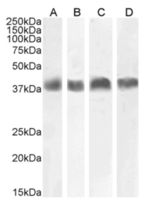
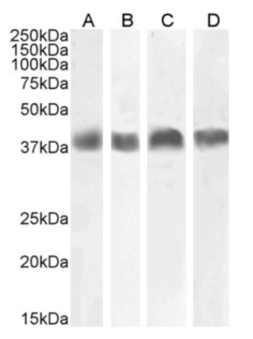
Western Blot using anti-Podoplanin (MAP tag) antibody PMab-1 (Ab00875-23.0) Mouse thymus (A), mouse brain (B), NIH3T3 cells (C) and mouse kidney (D) lysates (35µg protein in RIPA buffer) were resolved on a 10% SDS PAGE gel and blots probed with the chimeric rabbit IgG version of 1B4 (Ab01258-23.0) at 0.01 µg/ml, 0.1 µg/ml, 0.001 µg/ml and 0.003 µg/ml, respectively, before detection using an anti-rabbit secondary antibody. A primary incubation of 1h was used and protein was detected by chemiluminescence.
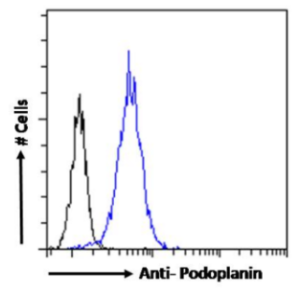
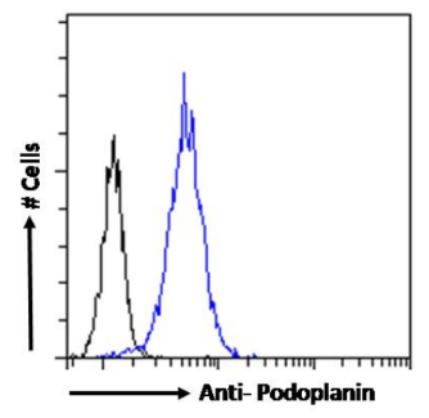
Flow-cytometry using the anti-Podoplanin (MAP tag) antibody PMab-1 (Ab00875) NIH3T3 cells were stained with anti-Fluorescein IgG antibody (4-4-20; isotype control, black line) or the rabbit IgG-chimeric version of PMab-1 (Ab00875-23.0, blue line) at a dilution of 1:100 for 1h at RT. After washing, bound antibody was detected using a goat anti-rabbit IgG AlexaFluor® 488 antibody at a dilution of 1:1000 and cells analyzed using a FACSCanto flow-cytometer.
ebiomall.com


>
>
>
>
>
>
>
>
>
>
>
>
抗体都是免疫球蛋白而免疫球蛋白不一定都是抗体。原因是抗体是由浆细胞产生,且能与相应抗原特异性结合发挥免疫功能的球蛋白。而免疫球蛋白是具有抗体活性或化学结构与抗体相似的球蛋白,如骨髓瘤患者血清中异常增高的骨髓瘤蛋白,是由浆细胞瘤产生,其结构与抗体相似,但无免疫功能。因此,免疫球蛋白可看做是化学结构上的概念,抗体则是生物学功能上的概念。
二、保持乐观情绪乐观的态度可以维持人体于一个最佳的状态,尤其是在现今社会,人们面临的压力很大,巨大的心理压力会导致对人体免疫系统有抑制作用的荷尔蒙成分增多,所以容易受到感冒或其它疾病的侵袭。
三、限制饮酒每天饮低度白酒不要超过100毫升,黄酒不要超过250毫升,啤酒不要超过1瓶,因为酒精对人体的每一部分都会产生消极影响。即使喝葡萄酒可以降低胆固醇,也应该限制每天一杯,过量饮用会给血液与心脏等器官造成很大破坏。
四、参加运动专家进行的3项研究指出,每天运动30到45分钟,每周5天,持续12周后,免疫细胞数目会增加,抵抗力也相对增加。运动只要心跳加速即可,晚餐后散步就很适合。
五、补充维生素每天适当补充维生素和矿物质。专家指出,身体抵抗外来侵害的武器,包括干扰素及各类免疫细胞的数量与活力都和维生素与矿物质有关。
六、改善体内生态环境用微生态制剂提高免疫力的研究和使用由来已久。研究表明,以肠道双歧杆菌、乳酸杆菌为代表的有益菌群具有广谱的免疫原性,能刺激负责人体免疫的淋巴细胞分裂繁殖,同时还能调动非特异性免疫系统,去“吃”掉包括病毒、细菌、衣原体等在内的各种可致病的外来微生物,产生多种抗体,提高人体免疫能力。对于健康人来说,不妨“食疗”,多吃些乳酸菌饮料;而健康边缘人群,可以用微生态制剂来调节体内微生态平衡。能提高免疫力的食品
1.灵芝:灵芝可增强人体的免疫力,这是因为灵芝含有抗癌效能的多糖体,此外,还含有丰富的锗元素。锗能加速身体的新陈代谢,延缓细胞的衰老,能通过诱导人体产生干扰素而发挥其抗癌作用;
2.新鲜萝卜:因其含有丰富的干扰素诱导剂而具有免疫作用;
3.人参蜂王浆:能提高机体免疫力及内分泌的调节能力,并含具有防癌作用的蜂乳酸;
4.蘑菇、猴头菇、草菇、黑木耳、银耳、车养、百合等:都有明显增强免疫力的作用;
5.香菇:香菇所含的香菇多糖能增强人体免疫力。展开