Cell culture contamination control: Detection
Detection and identification of Mycoplasma
Venor®GeM Classic Mycoplasma PCR Detection Kit
Venor®GeMMycoplasma PCR Detection Kit for rapid and reliable detection of mycoplasma in various in situ biologicals including cell cultures and virus stocks
Minerva Biolabs
| Catalogue No. | Description | Pack Size | Price | Qty |
|
|---|---|---|---|---|---|
| 11-1025 | Venor®GeM Classic Mycoplasma PCR Detection Kit | 25 tests | £125.00 | Quantity | Add to Order |
| 11-1050 | Venor®GeM Classic Mycoplasma PCR Detection Kit | 50 tests | £221.00 | Quantity | Add to Order |
| 11-1100 | Venor®GeM Classic Mycoplasma PCR Detection Kit | 100 tests | £387.00 | Quantity | Add to Order |
| 11-1250 | Venor®GeM Classic Mycoplasma PCR Detection Kit | 250 tests | £831.00 | Quantity | Add to Order |
| 11-1905 | Venor®GeM extra internal control DNA | 4 vials | £19.00 | Quantity | Add to Order |
Related products
Venor®GeM Classic Mycoplasma PCR Detection Kit
Venor®GeMMycoplasma PCR Detection Kit for rapid and reliable detection of mycoplasma in various in situ biologicals including cell cultures and virus stocks
Minerva Biolabs

Venor®GeM Classic
Venor®GeM Classic allows fast, reliable and time-saving routine monitoring of mycoplasma contamination by PCR.
Type of PCR
For conventional, endpoint PCR
Recommended Use / Scope
Applicable in research and industry for direct testing of cell cultures and biologicals. Intended for research use only. Not recommended for clinical diagnostics.
Kit Components
Lyophilized primer sets and nucleotides 10x reaction buffer optimized for MB Taq DNA Polymerase Lyophilized positive control DNA Lyophilized internal amplification control
Package Sizes
Primer sets and nucleotides are prepared in aliquots of 25 tests.
- Cat. No. 11-1025 25 Tests
- Cat. No. 11-1050 50 Tests
- Cat. No. 11-1100 100 Tests
- Cat. No. 11-1250 250 Tests
Result evaluation
Gel electrophoresis at endpoint of PCR
Required Consumables
Polymerase. We highly recommend our reliable hot-start MB Taq DNA Polymerase, Cat.-No. 53-0050/100/200/250. PCR reaction tubes
Optional for process validation and EP 2.6.7 compliant testing: Internal Control DNA extra (4 vials for 300 µl each of internal amplification control; Cat. No. 11-1905) 10CFU™ Sensitivity Standards available for all EP listed mycoplasma species
Required lab devices
Regular PCR cycler Agarose gel electrophoresis and DNA staining system Pipetting equipment Tube centrifuge
Shelf Life and Storage
Components are maintainable at2 to8 °C for at least 6 months. After rehydratisation the reagents must be stored at -18 °C.
EP 2.6.7 compliance
Use of Venor®GeM Classic for QA testing of biologicals like master and working cell banks, autologous cells, culture media, bulk harvest and final product testing according to EP 2.6.7 is applicable after appropriate sample preparation and process validation.
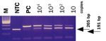
Fig. Amplified PCR products are visualized by standard gel electrophoresis.
If you cannot find the answer to your problem below then please contact us or telephone 01954 210 200
Venor®GeM Classic Mycoplasma PCR Detection Kit
Venor®GeMMycoplasma PCR Detection Kit for rapid and reliable detection of mycoplasma in various in situ biologicals including cell cultures and virus stocks
Minerva Biolabs
You can find your Venor®GeM Classic protocols here:
Click to download:
Venor®GeM Classic Kit Full Protocol
alternatively,
if you are in a rush click to view:
Venor®GeM Classic Kit Quick Visual Guide
If you cannot find the answer to your problem below then please contact us or telephone 01954 210 200
Venor®GeM Classic Mycoplasma PCR Detection Kit
Venor®GeMMycoplasma PCR Detection Kit for rapid and reliable detection of mycoplasma in various in situ biologicals including cell cultures and virus stocks
Minerva Biolabs
Venor®GeM Mycoplasma Detection Kits
Anugraham M. et al., (2014). Specific Glycosylation of Membrane Proteins in Epithelial Ovarian Cancer Cell Lines: Glycan Structures Reflect Gene Expression and DNA Methylation Status. Molecular & Cellular Proteomics, 13(9):2213-32. doi: 10.1074/mcp.M113.037085
Brinzeu D.G.T. et al., (2008). Microbial and Fungal Contamination of Keratinocyte and Fibroblast Cell Cultures. Journal of Experimental Medical & Surgical Research, 3 / 123 – 128.
Chal J. et al., (2016). Generation of human muscle fibers and satellite-like cells from human pluripotent stem cells in vitro. Nature Protocols 11(10):1833-50. doi: 10.1038/nprot.2016.110.
Dzieran J. et al., (2013). Comparative Analysis of TGF-β/Smad Signaling Dependent Cytostasis in Human Hepatocellular Carcinoma Cell Lines. PLoS One, 8(8):e72252. doi: 10.1371/journal.pone.0072252.
Falagan-Lotsch P.et al., (2015). Performance of PCR-based and bioluminescent assays for mycoplasma detection. Journal of Microbiological Methods, 118:31-6. doi: 10.1016/j.mimet.2015.08.010.
Gutzeit C.et al., (2014). Exosomes derived from Burkitt"s lymphoma cell lines induce proliferation, differentiation, and class-switch recombination in B cells. Journal of Immunology, 192(12):5852-62. doi: 10.4049/jimmunol.1302068.
Henrich B.et al., (2010). Mycoplasma salivarium detected in a microbial community with Candida glabrata in the biofilm of an occluded biliary stent. Journal of Medical Microbiology, 59, 239–241. doi: 10.1099/jmm.0.013110-0.
Loring J.F., Wesselschmidt R.L., and Schwartz P.H. (2007). Human Stem Cell Manual: A Laboratory Guide.Elsevier Inc.
Maass V.et al., (2011). Sequence homologies between Mycoplasma and Chlamydia spp. lead to false-positive results in chlamydial cell cultures tested for mycoplasma contamination with a commercial PCR assay. Journal of Clinical Microbiology, 49(10):3681-2. doi: 10.1128/JCM.01092-11.
Malenovska H. and Reichelova M., (2011). Elimination of mycoplasma contamination of virus stocks.Veterinarni Medicina, 56 (11): 547–550.
Mudduluru G.et al., (2015). A Systematic Approach to Defining the microRNA Landscape in Metastasis. Cancer Research, 75(15). doi: 10.1158/0008-5472.CAN-15-0997.
Nübling C.M.et al., (2015). World Health Organization International Standard to Harmonize Assays for Detection of Mycoplasma DNA. Applied and Environmental Microbiology, 81(17):5694-702. doi: 10.1128/AEM.01150-15.
Oksvold M.P.et al., (2012). Effect of cycloheximide on epidermal growth factor receptor trafficking and signaling. FEBS Letters, 586(20):3575-81. doi: 10.1016/j.febslet.2012.08.022.
Pandrangi S.L.et al., (2014). Establishment and characterization of two primary breast cancer cell lines from young Indian breast cancer patients: mutation analysis. Cancer Cell International, 14(1):14. doi: 10.1186/1475-2867-14-14.
Panosa C. et al., (2013). Development of an Epidermal Growth Factor Derivative with EGFR Blocking Activity. PLoS One, 8(7):e69325. doi: 10.1371/journal.pone.0069325.
Pérez-Garay M. et al., (2010). 2,3-Sialyltransferase ST3Gal III Modulates Pancreatic Cancer Cell Motility and Adhesion In Vitro. PLoS One, 5(9): e12524. doi: 10.1371/journal.pone.0012524.
Picco G. et al., (2017). Loss of AXIN1 drives acquired resistance to WNT pathway blockade in colorectal cancer cells carrying RSPO3 fusions. EMBO Molecular Medicine, 9(3):293-303. doi: 10.15252/emmm.201606773.
Pietrosi G. et al., (2015). Phases I–II Matched Case-Control Study of Human Fetal Liver Cell Transplantation for Treatment of Chronic Liver Disease. Cell Transplantation, 24(8):1627-38. doi: 10.3727/ 096368914X682422.
Pontarin G. et al., (2006). Mitochondrial DNA Depletion and Thymidine Phosphate Pool Dynamics in a Cellular Model of Mitochondrial Neurogastrointestinal Encephalomyopathy. The Journal of Biological Chemistry, 281(32):22720-8. doi: 10.1074/jbc.M604498200.
Rappa G. et al., (2015). Ethanol induces upregulation of the nerve growth factor receptor CD271 in human melanoma cells via nuclear factor-κB activation. Oncology Letters, 10: 815-821. doi: 10.3892/ol.2015.3343.
Sajadian S.O. et al., (2016). Vitamin C enhances epigenetic modifications induced by 5-azacytidine and cell cycle arrest in the hepatocellular carcinoma cell lines HLE and Huh7. Clinical Epigenetics, 8:46. doi: 10.1186/s13148-016-0213-6.
Samanta D. et al., (2012). Smoking attenuates Transforming growth factor-β-mediated tumor suppression function through down-regulation of Smad3 in lung cancer. Cancer Prevention Research, 5(3): 453–463. doi:10.1158/1940-6207.
Schmitt M. et al., (2009). High-throughput detection and multiplex identification of cell contaminations. Nucleic Acids Research, 37(18):e119. doi:10.1093/nar/gkp581.
Smith C. (2005). Trouble in the hood: culturing difficult cell types.Nature Methods 2, 385–391. doi: 10.1038/nmeth0505-385
Thies A. et al., (2007). Clinically proven markers of metastasis predict metastatic spread of human melanoma cells engrafted in scid mice. British Journal of Cancer, 96(4):609-16. doi: 10.1038/sj.bjc.6603594.
Vickovic S. et al., (2016). Massive and parallel expression profiling using microarrayed single-cell sequencing. Nature Communications, 7:13182. doi: 10.1038/ncomms13182.
Waddington R.J. and Sloan A.J. (2017). Tissue Engineering and Regeneration in Dentistry – Current Strategies.John Wiley & Sons, Ltd.
Winter S. et al., (2016). Methylomes of renal cell lines and tumors or metastases differ significantly with impact on pharmacogenes. Scientific Reports, 6:29930. doi: 10.1038/srep29930.
Wolff J. et al., (2013). GMP-level adipose stem cells combined with computer-aided manufacturing to reconstruct mandibular ameloblastoma resection defects: Experience with three cases. Annals of Maxillofacial Surgery, 3(2):114-25. doi: 10.4103/2231-0746.119216.
If you cannot find the answer to your problem below then please contact us or telephone 01954 210 200
Venor®GeM Classic Mycoplasma PCR Detection Kit
Venor®GeMMycoplasma PCR Detection Kit for rapid and reliable detection of mycoplasma in various in situ biologicals including cell cultures and virus stocks
Minerva Biolabs
Click here to download full product information.
If you cannot find the answer to your problem below then please contact us or telephone 01954 210 200
AthensResearch&Technology公司位于美国乔治亚州雅典市,从1986年以来一直致力于纯化高纯度,高活性的人类蛋白质和开发这些蛋白的多克隆抗血清。我们的常规产品包括丝氨酸蛋白酶、蛋白酶抑制剂、中性粒细胞酶、载脂蛋白、脂蛋白,血小板蛋白、转铁蛋白、免疫球蛋白等。二十年多年来,世界各地的研究人员都在使用我们公司的产品。在炎症、冠状动脉疾病、自身免疫性疾病、癌症、老年痴呆症等等研究的主要科学出版物中都能看到引用我们公司的产品。我们提供优质,可靠的产品和出色的客户服务。我们期待着您的业务。如果需要定制研究服务,AthensResearch&Technology公司能够根据您的项目要求提供无与伦比的试剂。
公司主要产品:一、凝血因子相关系列产品:包括多款从人血浆、血小板中分离纯化的凝血因子相关蛋白。二、酶及抑制剂系列产品:包括多款从人血浆、红细胞分离纯化的a-抗糜蛋白酶、a1/2抗胰蛋白酶、抗凝血酶、C1酯酶抑制因子、Calpain-1等酶及其抑制剂产品。三、免疫球蛋白系列产品:包括多款从人血浆、骨髓瘤、初乳以及狗和小鼠血浆中分离纯化的免疫球蛋白产品。四、脂蛋白及载脂蛋白系列产品:包括包括多款从人血浆中分离纯化的载脂蛋白及脂蛋白(高密度、中等密度、低密度、极低密度脂蛋白)产品。五、低内毒素蛋白系列产品:包括多款适宜于细胞培养的低内毒素脂蛋白、免疫球蛋白及转铁蛋白产品。六、乳蛋白系列产品:包括从人乳中分离纯化的乳白蛋白、乳铁传递蛋白及分泌型免疫球蛋白A。七、中性粒细胞源蛋白系列产品:包括多款纯化分离自人中性粒细胞的Azurocidin 、CathepsinG及Lactoferrin等蛋白产品。八、血小板相关系列产品:包括纯化分离自人血小板的血小板因子4及凝血酶致敏蛋白。九、转铁蛋白及脱铁转铁蛋白(APOTRANSFERRINS)系列产品:包括多款纯化分离自人、大小鼠、狗及食蟹猴血浆中的转铁蛋白/脱铁转铁蛋白,可应用于细胞或组织培养。十、抗体系列产品:包括多款兔、大鼠、羊源的多克隆抗体产品。
ebiomall.com