![absoluteantibody/Anti-F4/80 [Cl:A3-1 (recombinant version)]/200 μg/Ab00106-2.3](images/no_picture.gif)
UniProt Accession Number of Target Protein: Q61549
Alternative Name(s) of Target: EGF-like module; Cell surface glycoprotein F4/80; EGF-like module receptor 1; EMR1 receptor; Em Gpf4; TM7; Ly-71; f480; f-480; f4 80; f 480; f-4/80; f4-80Immunogen: Thioglycollate stimulated peritoneal macrophages of mouse origin.
Specificity: This antibody recognises the mouse F4/80 antigen, a 160kD glycoprotein, expressed by murine macrophages.
Application Notes: The anti-F4/80 antibody, clone Cl:A3-1, has been very widely used in flow cytometry, microscopy and Western blotting since its generation by Austyn and Gordon in 1980. We are the only supplier of recombinant versions of Cl:A3-1 which, whilst having the same variable domains as the original, has been shown to block hybridoma derived anti-F4/80:APC binding to in vitro generated bone marrow-derived (BMDMs) and ex vivo splenic murine macrophages. This was tested using both the recombinant version of the original isotype (rat IgG2b) as well as a murinised version (IgG2a, Ab00106-2.0). Further testing on BMDMs shows that our Fc Silent™ rat IgG2b is a valuable new reagent which shows distinct labelling of F4/80-positive cell populations, whilst the isotype control shows no binding, suggesting that only the antigen binding domains are involved in binding, promoting precision of labelling. The murinised versions will be valuable tools in the study of the role of the murine macrophage in vivo without the artefacts rat-derived antigen being co-administered may harbour. Particularly the murine IgG2a (Ab00106-2.0) should be an excellent candidate for macrophage depletion studies, with the Fc Silent™ (Ab00106-2.3) version being the ideal negative control. Using our recombinant production platform, any species and isotype are possible and our Support Team (support@absoluteantibody.com) will be glad to help you with finding the right variant to match your experimental needs.
Antibody first published in:Austyn JM, Gordon S.F4/80, a monoclonal antibody directed specifically against the mouse macrophage.Eur J Immunol. 1981 Oct;11(10):805-15.PMID:7308288Note on publication:Describes the making of the antibody and its specificity for macrophages (Flow Cytometry) and proposes its potential use as a marker.
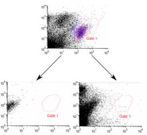
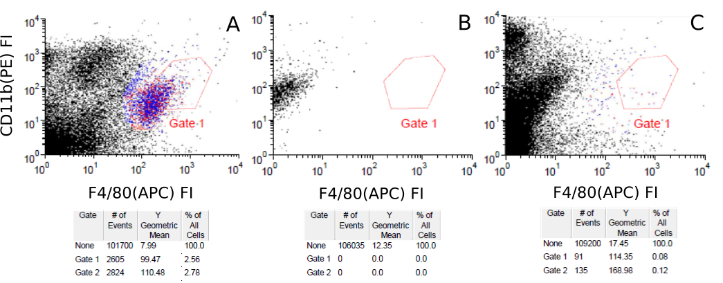
Competitive flow-cytometry assay between Absolute Antibody anti-F4/80 (Ab00106) CI:A3-1 variants and an existing commercial anti-F4/80 antibody. Mouse (Mus musculus) splenocytes were labelled ex vivo with a commercially available APC-labelled anti-F4/80 antibody and APE labelled anti-CD11b antibody and subject to flow-cytometry analysis (A), in which a small subpopulation of F4/80-CD11b positive cells may be observed. Subsets of commerical anti-F4/80 antibody-labelled splenocytes were also subsequently incubated with unlabelled versions of either the rat ( Rattus norvegicus ) IgG2b chimeric version (Ab00106-8.1, B) or mouse IgG2A chimeric (Ab00106-2.0, C) version of CI:A3-1. Loss of the F4/80-CD11b positive subpopulation may be observed, demonstrating displacement of the commercial antibody and the specificity of CI:A3-1.
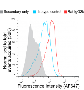
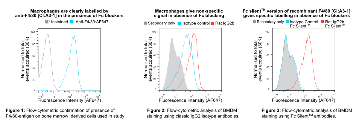
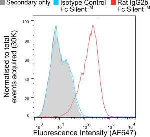
In Figure 1 murine bone marrow-derived macrophages (BMDMs) were pre-blocked with rat anti-mouse CD16 & CD32 (clone FCR-4G8) and stained with non-recombinant anti-F4/80 [Cl:A3-1] conjugated to Alexa Fluor® 647 (AF647), all commercially available from competitors. InFigure 2 BMDMs were stained with recombinant anti-F4/80 [Cl:A3-1] or isotype control (anti-fluorescein [4-4-20 (enhanced)] IgG2b (Ab00102-8.1). InFigure 3 BMDMs were stained with Fc Silent™ recombinant anti-F4/80 [Cl:A3-1] (Ab00106-8.4) or isotype control (Fc Silent™ anti-fluorescein [4-4-20 (enhanced)] IgG2b (Ab00102-8.4). These were fluorescently labelled using the secondary antibody, goat IgG anti-rat IgG (H&L-chain) polyclonal antibody directly conjugated to Alexa Fluor® 647(AF647) commercially available from a competitor. Whilst inFigure 2 the highest fluorescence signal is seen with the recombinant anti-F4/80 IgG2 (the isotype of the original hybridoma-derived antibody), the isotype control IgG2b (Ab00102-8.1) shows considerable signal overlap, indicative of binding of the antibody to Fc-receptors. This illustrates the importance of isotype controls in such experiments when using conventional antibody formats particularly when Fc-blocking reagents are incompatible with the system used due to reactivity with the secondary antibody. The Fc silent™ format however overcomes this issue as seen inFigure 3, where the Fc silent™ recombinant anti-F4/80 (Ab00102-8.4) yields a strong and distinct signal, whilst the isotype control (Ab00102-8.4) shows no discernible difference to the background staining from the secondary antibody alone. Therefore, with Fc Silent™ reagents, no Fc-blocking productsare required. [Data courtesy of Lewis Taylor.]

In Figure 1 murine bone marrow-derived macrophages (BMDMs) were pre-blocked with rat anti-mouse CD16 & CD32 (clone FCR-4G8) and stained with non-recombinant anti-F4/80 [Cl:A3-1] conjugated to Alexa Fluor® 647 (AF647), all commercially available from competitors. InFigure 2 BMDMs were stained with recombinant anti-F4/80 [Cl:A3-1] or isotype control (anti-fluorescein [4-4-20 (enhanced)] IgG2b (Ab00102-8.1). InFigure 3 BMDMs were stained with Fc Silent™ recombinant anti-F4/80 [Cl:A3-1] (Ab00106-8.4) or isotype control (Fc Silent™ anti-fluorescein [4-4-20 (enhanced)] IgG2b (Ab00102-8.4). These were fluorescently labelled using the secondary antibody, goat IgG anti-rat IgG (H&L-chain) polyclonal antibody directly conjugated to Alexa Fluor® 647(AF647) commercially available from a competitor. Whilst inFigure 2 the highest fluorescence signal is seen with the recombinant anti-F4/80 IgG2 (the isotype of the original hybridoma-derived antibody), the isotype control IgG2b (Ab00102-8.1) shows considerable signal overlap, indicative of binding of the antibody to Fc-receptors. This illustrates the importance of isotype controls in such experiments when using conventional antibody formats particularly when Fc-blocking reagents are incompatible with the system used due to reactivity with the secondary antibody. The Fc silent™ format however overcomes this issue as seen inFigure 3, where the Fc silent™ recombinant anti-F4/80 (Ab00102-8.4) yields a strong and distinct signal, whilst the isotype control (Ab00102-8.4) shows no discernible difference to the background staining from the secondary antibody alone. Therefore, with Fc Silent™ reagents, no Fc-blocking productsare required. [Data courtesy of Lewis Taylor.]
ebiomall.com


>
>
>
>
>
>
>
>
>
>
>
拼音名:Chunhuashui
英文名:PurifiedWater
【性状】本品为无色的澄清液体;无臭,无味。
【检查】酸碱度取本品10ml,加甲基红指示液2滴,不得显红色;另取10ml,加溴麝香草酚蓝指示液5滴,不得显蓝色。氯化物、流酸盐与钙盐取本品,分置三支试管中,每管各50ml。第一管中加硝酸5滴与硝酸银试液1ml,第二管中加氯化钡试液2ml,第三管中加草酸铵试液2ml,均不得发生浑浊。
硝酸盐取本品5ml置试管中,于冰浴中冷却,加10%氯化钾溶液0.4ml与0.1%二苯胺硫酸溶液0.1ml,摇匀,缓缓滴加硫酸5ml,摇匀,将试管子50℃水浴中放置15分钟,溶液产生的蓝色与标准硝酸盐溶液[取硝酸钾0.163g,加水溶解并稀释至100ml,摇匀,精密量取1ml,加水稀释成100ml,再精密量取10ml,加水稀释成100ml,摇匀,即得(每1ml相当于1pgNO3)0.3ml,加无硝酸盐的水4.7ml,用同一方法处理后的颜色比较,不得更深(0.000006%)。
亚硝酸盐取本品10ml,置纳氏管中,加对氨基苯磺酰胺的稀盐酸溶液(1→100)lml与盐酸菜乙H肢溶液(0.l+100)1ml,产生的粉红色,与标准亚硝酸盐溶液〔取亚硝酸钠0.750g(按干燥品计算),加水溶解,稀释至100ml,摇匀,精密量取1ml,加水稀释成100ml,摇匀,再精密量取1ml,加水稀释成50ml,摇匀,即得(每1ml相当于1μgNO2)]0.2ml,加无亚硝酸盐的水9.8ml,用同一方法处理后的颜色比较,不得更深(0.000002%)。
氨取本品50ml,加碱性碘化汞钾试液2ml,放置15分钟;如显色,与氯化铵溶液(取氯化铵31.5mg,加无氨水适量使溶解并稀释成1000ml)1.5ml,加元氨水48ml与碱性碘化汞钾试液2ml制成的对照液比较,不得更深(0.00003%)。
二氧化碳取本品25ml,置50ml具塞量筒中,加氢氧化钙试液25ml,密塞振摇,放置,小时内不得发生浑浊。
易氧化物取本品100ml,加稀硫酸10ml,煮沸后,加高锰酸钾滴定液(0.02mol/L)0.10ml,再煮沸10分钟,粉红色不得完全消失。
不挥发物取本品100ml,置105℃恒重的蒸发皿中,在水浴上蒸干,并在105℃干燥至恒重,遗留残渣不得过1mg。
重金属取本品50ml,加水18.5ml,蒸发至20ml,放冷,加醋酸盐缓冲液(pH3.5)2ml与水适量使成25ml,加硫代乙酰胺试液2ml,摇匀,放置2分钟,与标准铅溶液1.5ml加水18.5ml用同一方法处理后的颜色比较,不得更深(0.00003%)。
微生物限度取本品,采用薄膜过滤法处理后,依法检查(附录ⅪJ),细菌、霉菌和酵母菌总数每1ml不得过100个。
【贮藏】密闭保存。
【化学成分】本品为蒸馏法、离子交换法、反渗透法或其他适宜的方法制得的供药用的水,不含任何附加剂。
【分子式与分子量】H2O18.02
【药理作用】溶剂、稀释剂
这里药典纯化水标准中并无PH值项目,请问对纯化水有PH值的要求吗,范围应在多少?请说明出处?
在纯化水检测中,检验酸碱度合格,但是发现PH在8左右。如果按以上标准检验合格,是否要考虑PH值?请知道的解答,谢谢!
:)
我在做一细菌不同酸碱度生长状况时,发现这些奇怪现象:pH=3的培养基灭菌(TSB液体培养基)灭菌后pH上升到到9.2!而原来pH=9.0的降到8.7(基本没多少变化),请问各位大侠,这是什么原因?
一般做不同酸碱度生长实验时,该如何才能防止pH在湿热灭菌后基本不变化?
是否可以理解为纯化水得PH范围为6.3-7.6?能否直接用pH计测量?谢谢!
1、弱酸和它的盐(如:HAc---NaAc)的水溶液组成;
2、弱碱和它的盐(如:NH3·H2O---NH4Cl)的水溶液组成;
3、多元弱酸的酸式盐及其对应的次级盐(如:NaH2PO4---Na2HPO4)的水溶液组成。
酸碱缓冲溶液的选型一般应根据具体情况进行选择。缓冲酸性可选用碱性缓冲液,缓冲酸性可采用碱性缓冲液。常用作缓冲溶液的酸类由弱酸及其共轭酸盐组合成的溶液具有缓冲作用。生化实验室常用的缓冲系主要有磷酸、柠檬酸、碳酸、醋酸、巴比妥酸、Tris(三羟甲基氨基甲烷)等系统,生化实验或研究工作中要慎重地选择缓冲体系,因为有时影响实验结果的因素并不是缓冲液的pH值,而是缓冲液中的某种离子。如硼酸盐、柠檬酸盐、磷酸盐和三羟甲基甲烷等缓冲剂都可能产生不需要的化学反应。
【酸碱缓冲溶液】由弱酸及其盐、弱碱及其盐组成的混合溶液,能在一定程度上抵消、减轻外加强酸或强碱对溶液酸碱度的影响,从而保持溶液的pH值相对稳定。这种溶液称为酸碱缓冲溶液。
由弱酸及其盐、弱碱及其盐组成的混合溶液,能在一定程度上抵消、减轻外加强酸或强碱对溶液酸碱度的影响,从而保持溶液的pH值相对稳定。这种溶液称为缓冲溶液。
两个CEX方法A和B测定同一单抗,结果碱性峰比例差不多,酸性峰比例相差约7%,相应主峰也差了7%左右。
具体来说,A方法酸性峰高,主峰低,碱性峰稍微低点;B方法酸性峰低,主峰高,碱性峰稍微高点;另外也做了CIEF,结果呢和A方法更接近。
仔细比较起来,AB两个方法的峰性和数量差不多,就不知道为什么会有这么大的差异。两个方法一个用的WCX柱-磷酸缓冲液,一个用SCX柱-MES缓冲液
大家帮我分析下:
1.两个方法哪个方法更准确,是以酸性峰高的为准还是什么?为什么?
2.这显著差异是由方法造成,具体原因是什么?柱子?
3.CIEF的结果和A方法更接近,是不是可以由此证明A方法更好或者CIEF的方法更好(因为CIEF更快更方便)?
欢迎讨论~
纠正下,A方法用的是Tosoh的柱子,B方法用的是SCX柱。TOSOH的柱子是7um的填料,10cm长。SCX是10um的填料。我本人TOSOH的阳离子柱子用的很少,这次信手用用,结果发现差异很大
那我现在就考虑,在以后方法开发过程中,除了通过流动相pH和组成、梯度、柱子选择来获得样品主峰和酸碱性的最大分离,还要关注各峰比例。因为之前比较方法好坏都只看分离度,尤其是主峰和邻近峰的分离度,获得最大分离度,自然可以做到主峰尽可能纯,但从未认真比较过各峰比例。这是一个大疏忽吧!
另外,CIEF和CEX方法原理还是有点差异的,所以分的是不同的异质体,原液放行两个方法肯定是都要做的。问题就是在早期细胞株筛选和工艺开发阶段,哪个方法才是又快又准。CIEF(iCE280)一般15分钟一个样,比CEX快多了。如果CIEF测得主峰要低于CEX结果,是不是真的完全可以取代CEX呢?CEX分离出的峰远比CIEF的多!
欢迎大家继续讨论~
这就是说不用酸碱预处理吗?
Whatman的网站上没有DE52最大耐受压力,请问又经验的战友应该是多少?
Whatman的网站上:
DE32DryMicrogranularDEAECellulose
SimilarperformancecharacteristicsafterprecyclingasDE52.
DE52PreswollenMicrogranularDEAECellulose
ProbablythemostwidelyusedDEAEcelluloseintheworld;usedforbiopolymerswithlowtohighnegativecharges;exhibitsexcellentresolutionwithgoodflowrates.
附件是一本图书(MethodsinMolecularMedicine,)的章节,上面说:
WhatmanDEAE52comesalreadypreswollenandonlyneedstobetransferred
totherunningbuffer50mMTE8.
lAntibodiesUsingIonExchangeChromatography.pdf(87.06k)
pH(1)=pKa+lg[c(CH₃COONa)/c(CH₃COOH)]=pKa=4.74
通HCl后,溶液是c(CH₃COOH)=0.2mol/L、c(NaCl)=0.1mol/L的混合溶液,溶液pH按照弱酸溶液pH的求法求.
c(H⁺)=√[Ka*c(CH₃COOH)]=√(10^-4.74*0.2)=0.00191(mol/L)(采用了近似公式)
pH(2)=-lg{c(H⁺)}=2.72
两个pH求得,那么pH的变化量也就可得了.pH的变化量=|pH(2)-pH(1)|=|2.72-4.74|=2.02
1)PH缓冲溶液作用原理和pH值
当往某些溶液中加入一定量的酸和碱时,有阻碍溶液pH变化的作用,称为缓冲作用,这样的溶液叫做缓冲溶液.弱酸及其盐的混合溶液(如HAc与NaAc),弱碱及其盐的混合溶液(如NH3·H2O与NH4Cl)等都是缓冲溶液.
由弱酸HA及其盐NaA所组成的缓冲溶液对酸的缓冲作用,是由于溶液中存在足够量的碱A-的缘故.当向这种溶液中加入一定量的强酸时,H离子基本上被A-离子消耗:
所以溶液的pH值几乎不变;当加入一定量强碱时,溶液中存在的弱酸HA消耗OH-离子而阻碍pH的变化.
2)PH缓冲溶液的缓冲能力
在缓冲溶液中加入少量强酸或强碱,其溶液pH值变化不大,但若加入酸,碱的量多时,缓冲溶液就失去了它的缓冲作用.这说明它的缓冲能力是有一定限度的.
缓冲溶液的缓冲能力与组成缓冲溶液的组分浓度有关.0.1mol·L-1HAc和0.1mol·L-1NaAc组成的缓冲溶液,比0.01mol·L-1HAc和0.01mol·L-1NaAc的缓冲溶液缓冲能力大.关于这一点通过计算便可证实.但缓冲溶液组分的浓度不能太大,否则,不能忽视离子间的作用.
组成缓冲溶液的两组分的比值不为1∶1时,缓冲作用减小,缓冲能力降低,当c(盐)/c(酸)为1∶1时△pH最小,缓冲能力大.不论对于酸或碱都有较大的缓冲作用.缓冲溶液的pH值可用下式计算:
此时缓冲能力大.缓冲组分的比值离1∶1愈远,缓冲能力愈小,甚至不能起缓冲作用.对于任何缓冲体系,存在有效缓冲范围,这个范围大致在pKaφ(或pKbφ)两侧各一个pH单位之内.
弱酸及其盐(弱酸及其共轭碱)体系pH=pKaφ±1
弱碱及其盐(弱碱及其共轭酸)体系pOH=pKbφ±1
例如HAc的pKaφ为4.76,所以用HAc和NaAc适宜于配制pH为3.76~5.76的缓冲溶液,在这个范围内有较大的缓冲作用.配制pH=4.76的缓冲溶液时缓冲能力最大,此时(c(HAc)/c(NaAc)=1.
3)PH缓冲溶液的配制和应用
为了配制一定pH的缓冲溶液,首先选定一个弱酸,它的pKaφ尽可能接近所需配制的缓冲溶液的pH值,然后计算酸与碱的浓度比,根据此浓度比便可配制所需缓冲溶液.
以上主要以弱酸及其盐组成的缓冲溶液为例说明它的作用原理、pH计算和配制方法.对于弱碱及其盐组成的缓冲溶液可采用相同的方法.
PH缓冲溶液在物质分离和成分分析等方面应用广泛,如鉴定Mg2离子时,可用下面的反应:
白色磷酸铵镁沉淀溶于酸,故反应需在碱性溶液中进行,但碱性太强,可能生成白色Mg(OH)2沉淀,所以反应的pH值需控制在一定范围内,因此利用NH3·H2O和NH4Cl组成的缓冲溶液,保持溶液的pH值条件下,进行上述反应.