- Extract high quality & purity total RNA including miRNA
- No phenol step required; isolate all RNA in one fraction
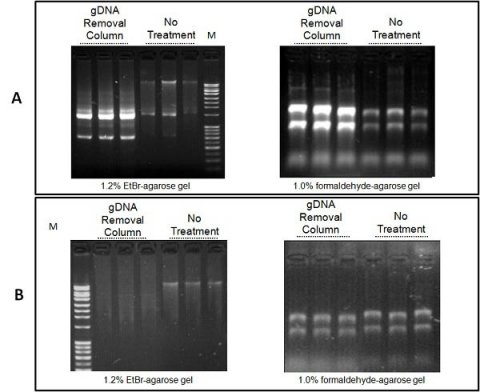
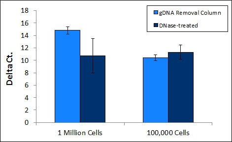
- Genomic DNA Removal Column for efficient elimination of gDNA
- Bind & elute all RNA irrespective of size or GC content, without bias
- Efficiently extract small RNA irrespective of GC content
- Very sensitive & linear down to a few cells without the need for carrier RNA
- Convenient & fast spin column format
- Isolate from a wide variety of specimens
- Buffer chemistry inactivates viruses including SARS-CoV-2 - Explore the Application Note
Total RNA Purification Plus Kit
This kit purifies total RNA including miRNA from biological samples and employs an extra column for the efficient removal of contaminating gDNA, thereby replacing the enzymantic DNase step.
Efficiently extract total RNA from a range of samples including cells, bacteria, yeast, virus and bodily fluids including plasma/serum, blood, saliva, CSF and more. Extract high quality and purity RNA with excellent RIN values and A260/A280 suitable for downstream applications including qRT-PCR, RT-PCR, microarrays, NGS and more.
The kit purifies all sizes of RNA from large mRNA, lncRNA down to microRNA (miRNA) in the same fraction without the requirement of phenol. Isolate all RNA sequences at an equal rate irrespective of size. Moreover, when the RNA sequences are small (e.g. miRNA), the column binds small RNAs regardless of their GC content.
ebiomall.com


>
>
>
>
>
>
>
>
>
>
>
>
一染色程序
1.石蜡切片HE染色(常规HE染色)
(1)二甲苯Ⅰ 5~10min
(2)二甲苯Ⅱ 5~10min
(3)无水乙醇Ⅰ 1~3min
(4)无水乙醇Ⅱ 1~3min
(5)95%乙醇Ⅰ 1~3min
(6)95%乙醇Ⅱ 1~3min
(7)80%乙醇 1min
(8)蒸馏水 1min
(9)苏木素液染色 5~10min
(10)流水洗去苏木素液 1min
(11)1%盐酸-乙醇 1~3s
(12)稍水洗 1~2s
(13)返蓝(用温水或1%氨水等) 5~10s
(14)流水冲洗 1~2min
(15)蒸馏水洗 1~2min
(16)0.5%伊红液染色 1~3min
(17)蒸馏水稍洗 1~2s
(18)80%乙醇 1~2s
(19)95%乙醇Ⅰ 2~3min
(20)95%乙醇Ⅱ 2~3min
(21)无水乙醇Ⅰ &, nbsp; 3~5min
(22)无水乙醇Ⅱ 3~5min
(23)石炭酸-二甲苯 3~5min
(24)二甲苯Ⅰ 3~5min
(25)二甲苯Ⅱ 3~5min
(26)二甲苯Ⅲ 3~5min
(27)中性树胶封固
注:①(12)和(13)项可省去,但(14)的冲水时间需延长至10~15min(细胞核显示更清晰)。
②(23)项可用无水乙醇代替;北方地区可省略。
2.冰冻切片HE染色
(1)冰冻切片固定 10~30s
(2)稍水洗 1~2s
(3)苏木素液染色(60℃) 30~60s
(4)流水洗去苏木素液 5~10s
(5)1%盐酸-乙醇 &n, bsp; &nbs, p; &nb, sp; 1~3s
(6)稍水洗 1~2min
(7)返蓝(用温水或1%氨水等) 5~10s
(8)流水冲洗 15~30s
(9)0.5%伊红液染色 1~2min
(10)蒸馏水稍洗 1~2min
(11)80%乙醇 1~2min
(12)95%乙醇 1~2min
(13)无水乙醇Ⅰ 1~2min
(14)无水乙醇Ⅱ 1~2min
(15)石炭酸-二甲苯 2~3min
(16)二甲苯Ⅰ 2~3min
(17)二甲苯Ⅱ 2~3min
(18)中性树胶封固
注:①(7)和(8)项可省去,但(9)的冲水时间需延长至10~15min(细胞核显示更清晰)。
②(15)项可用无水乙醇代替;北方地区可省略。
二染色结果:细胞核呈蓝色,细胞浆、肌纤维、胶原纤维和红细胞呈不同程度的红色。钙盐和细菌可呈蓝色或紫蓝色。
三染色注意事项
1.切片染色前,应彻底脱蜡。
2.用含有升汞液体固定的组织,其切片染色前应先脱去汞盐:
(1)石蜡切片脱蜡至水洗
(2)Lugol液 20min
(3)流水冲洗 5min
(4)95%乙醇 10min
(5)水洗 1min
(6)5%次亚硫酸钠水溶液 5min
(7)流水冲洗 5min
(8)显微镜观察除汞满意后,转入HE染色
3.脱除福尔马林色素(必要时):
(1)石蜡切片脱蜡至水洗
(2)1%NaOH(1ml)与80%乙醇(99ml)混合液 10min
(3)流水冲洗 5min
(4)转入HE染色
4.严格执行HE染色流程,用显微镜控制细胞核的苏木素染色质量。HE染片应着色鲜艳,红、蓝分明,对比清晰。
5.载玻片自二甲苯中取出后,应立即用洁净、光亮的盖玻片和稠度适宜的中性树胶湿封载玻片。封盖处内无气泡,外无溢胶。封片时必须进行操作人员和局部环境的二甲苯污染防护。不应将组织切片烤干或风干后再行封片。
6.必须在载玻片的一端牢贴标签。标签上应印有病理科所在的医院名称。标签上应清楚显示有关的病理号及其亚号;标签上的病理号应准确无误,无涂改。
7.制片完成后,技术人员应对切片与其相应的病理学检查记录单或取材工作记录单认真进行核对;确认无误后,将制备好的切片连同相关的活检申请单/活检记录单以及取材工作单等一并移交给有关的病理医师;交接双方经核对无误后,办理移交签字手续。
8.石蜡切片-HE染色的优良率(甲、乙级切片所占的比率)应≥85%。石蜡切片-HE染色质量的基本标准列于附表。
9.制片工作一般应在取材后2个工作日内完成(不含进行脱钙、脱脂等需要特殊处理的标本)。
10.制片过程出现意外情况时,技术室人员应及时向病理医师和科主任报告,设法予以补救。
附表 常规石蜡包埋-HE染色切片质量的基本标准
优 质 标 准 满 分 质 量 缺 陷 减 分
⒈组织切面完整, ①组织稍不完整:减1~3分 ②不完整:减4~10分
内镜咬检、穿刺标本切面数 10 ③未达到规定面数:减5分
⒉切片薄(3~5μm),厚薄均匀 10 ①切片厚(细胞重叠),影响诊断:减6~10分
②厚薄不均匀:减3~5分
⒊切片无刀痕、裂隙、颤痕 10 ①有刀痕、裂隙、颤痕,尚不影响诊断:减2分
②有刀痕、裂隙、颤痕,影响诊断:减5分
⒋切片平坦,无皱褶、折叠 10 ①有皱褶或折叠,尚不影响诊断:各减2分
②有皱褶折或折叠,影响诊断:各减5分
⒌切片无污染 10 有污染:减10分
⒍无气泡(切片与载玻片间/ 10 ①有气泡:减3分
盖片与切片、载玻片间), ②胶液外溢:减3分
盖片周围无胶液外溢
⒎透明度好 10 ①透明度差:减1~3分
②组织结构模糊:减5~7分
⒏细胞核与细胞浆染色对比清晰 10 ①细胞核着色灰淡或过蓝:减5分
②红(细胞浆)与蓝(细胞核)对比不清晰:减5分
⒐切片无松散,裱贴位置适当 10 ①切片松散:减5分
②切片裱贴位置不当:减5分
⒑切片整洁, ①切片不整洁:减3分
标签端正粘牢,编号清晰 10 ②标签粘贴不牢:减3分
③编号不清晰:减4分
合 计 100
切片质量分级标准: ①甲级片: ≥90分 (优)
②乙级片:75~89分(良)
③丙级片:60~74分(基本合格)
④丁级片: ≤59分 (不合格)
四HE染色试剂的配制
1.苏木素染液
(1)Harri苏木素染液
苏木素 1g
无水乙醇 10ml
硫酸铝钾 20g
蒸馏水 200ml
氧化汞 0.5g
冰醋酸 8ml
先用无水乙醇溶解苏木素,用蒸馏水加热溶解硫酸铝钾;然后将该两液合并煮沸,加入氧化汞,继续加热和搅拌溶液至深紫色,随即用冰水冷却,恢复至室温后过滤备用。使用前加入冰醋酸并混匀、过滤。
(2)Gill改良苏木素液
苏木素 2g
无水乙醇 250ml
硫酸铝钾 17g
蒸馏水 750ml
碘酸钠 0.2g
冰醋酸 20ml
先用无水乙醇溶解苏木素,用蒸馏水溶解硫酸铝钾;然后将该两液混合,再依次加入碘酸钠和冰醋酸。使用前过滤。
(3)Mayer改良苏木素液
A液:苏木素 2g
无水乙醇 40ml
B液:硫酸铝钾 100g
蒸馏水 600ml
将苏木素溶于无水乙醇中(A液);稍加热,使硫酸铝钾溶于蒸馏水中(B液)。将A液与B液混合后煮沸2min,再用蒸馏水补足至600 ml,加入400mg碘化钠充分混匀。染液呈紫红色。
2. 盐酸-乙醇分化液
浓盐酸 1 ml
70%乙醇 99 ml
3.伊红液
(1)0.25~0.5%伊红Y水溶液
伊红Y 0.25~0.5 g
蒸馏水 100 ml
冰醋酸 1滴
(2)0.5%伊红Y-氯化钙水溶液
伊红Y 0.5 g
蒸馏水 100 ml
无水氯化钙 0.5 g
(3)0.25~0.5%伊红Y-乙醇溶液。
伊红Y 0.25~0.5 g
80%乙醇 100 ml
4.石炭酸-二甲苯混合液
石炭酸 1份
二甲苯 3份
石蜡组织切片的HE染色
1.取材组织块,经固定后,常规石蜡包埋,4μm切片。
2.切片常规用二甲苯脱蜡,经各级乙醇至水洗:二甲苯(I)5min→二甲苯(Ⅱ)5min→100%乙醇2min→95%的乙醇1min→80%乙醇1min→75%乙醇1min→蒸馏水洗2min。
3.苏木素染色5min,自来水冲洗。
4.盐酸乙醇分化30s(提插数下)。
5.自来水浸泡15min或温水(约50℃)5min。
6.置伊红液2min。
7.常规脱水,透明,封片:95%乙醇(I)min→95%乙醇(Ⅱ)1min→100%乙醇(I)1min→100%乙醇(Ⅱ)1min→二甲苯石碳酸(3:1)1min→二甲苯(I)1min→二甲苯(Ⅱ)1min→中性树脂封固。
免疫组化操作规程
(一)、仪器设备
1)18cm不锈钢高压锅或电炉或医用微波炉;
2)水浴锅
(二)、试剂
1)PBS缓冲液(pH7.2~7.4):NaCl 137mmol/L,KCl 2.7mmol/L,Na2HPO4 4.3mmol/L, KH2PO4 1.4mmol/L。
2)0.01mol/L柠檬酸盐缓冲液(CB,pH6.0,1000ml):柠檬酸三钠 3g,柠檬酸 0.4g。
3)0.5mol/L EDTA缓冲液(pH8.0):700ml水中溶解186.1gEDTA·2H2O,用10 mmol/L NaOH调至pH8.0,加水至1000ml。
4)1mol/L的TBS缓冲液(pH8.0):在800ml水中溶解121gTris碱,用1N的HCl调至pH8.0,加水至1000ml。
5)酶消化液: a、0.1%胰蛋白酶液:用0.1%CaCl2(pH7.8) 配制。 b、0.4%胃蛋白酶液:用0.1N的HCl配制。
6)3%甲醇-H2O2溶液:用30%H2O2和80%甲醇溶液配制。
7)封裱剂:
a、甘油和0.5mmol/L碳酸盐缓冲液(pH9.0~9.5)等量混合;
b、油和TBS(或PBS)配制。
8)TBS/PBS pH9.0~9.5,适用于荧光显微镜标本;pH7.0~7.4适合于光学显微镜标本。
(三)、操作流程
1、脱蜡和水化
脱蜡前,应将组织芯片在室温中放置60分钟或60℃恒温箱中烘烤20分钟。
1)组织芯片置于二甲苯中浸泡10分钟,更换二甲苯后再浸泡10分钟;
2)无水乙醇中浸泡5分钟;
3)95%乙醇中浸泡5分钟;
4)70%乙醇中浸泡5分钟;
2、抗原修复
用于福尔马林固定的石蜡包埋组织芯片。
1)抗原热修复
(1)高压热修复 在沸水中加入EDTA(pH8.0)或0.01M枸橼酸钠缓冲溶液(pH6.0)。盖上不锈钢高压锅的盖子,但不进行锁定。将玻片置于金属染色架上,缓慢加压,使玻片在缓冲液中浸泡5分钟,然后将盖子锁定,小阀门将会升起来。10分钟后,去除热源,置入凉水中,当小阀门沉下去后打开盖子。本方法适用于较难检测或核抗原的抗原修复。
(2)煮沸热修复 电炉或者水浴锅加热0.01M枸橼酸钠缓冲溶液(pH6.0)至95℃左右,放入组织芯片加热10~15分钟。
(3)微波热修复 在微波炉里加热0.01M枸橼酸钠缓冲溶液(pH6.0)至沸腾后将组织芯片放入,断电,间隔5~10分钟,反复1-2次。适用的抗原有:AR,Bax,Bcl-2,C-fos,X-jun,C-kit,C-myc,E-cadherin,Chromogranin A,Cyclin,ER,Heat shock protein,HPV,Ki-67,MDMZ,p53,p34,p16,p15,P-glycoprotein,PKC,PR,PCNA,ras,Rb,TopoismeraseⅡ等。
2)酶消化方法 常用0.1%胰蛋白酶和0.4%胃蛋白酶液。胰蛋白酶使用前预热至37℃,切片也预热至37℃,消化时间约为5~30分钟;胃蛋白酶消化37℃时间为30分钟。适用于被固定遮避的抗原,其中有:Collagen,Complement,Cytokeratin,C-erB-2,GFAP,LCA,LN等。
3、免疫组织化学染色
SP法
1)脱蜡、水化;
2)PBS洗2~3次各5分钟;
3)3%H2O2(80%甲醇)滴加在TMA上,室温静置10分钟;
4)PBS洗2~3次各5分钟;
5)抗原修复;
6)PBS洗2~3次各5分钟;
7)滴加正常山羊血清封闭液,室温20分钟。甩去多余液体。
8)滴加Ⅰ抗50μl,室温静置1小时或者4℃过夜或者37℃1小时。
9)4℃过夜后需在37℃复温45分钟。
10)PBS洗3次各5分钟;
11)滴加Ⅱ抗40~50μl,室温静置,或37℃1小时;
12)II抗中可加入0.05%的tween-20。
13)PBS洗3次各5分钟;
14)DAB显色5~10分钟,在显微镜下掌握染色程度;
15)PBS或自来水冲洗10分钟;
16)苏木精复染2分钟,盐酸酒精分化;
17)自来水冲洗10~15分钟;
18)脱水、透明、封片、镜检。
SABC法
1)脱蜡、水化。
2)PBS洗两次各5分钟。
3)用蒸馏水或PBS配置新鲜的3%H2O2,室温封闭5~10分钟,蒸馏水洗3次。
4)抗原修复。
5)PBS洗5分钟。
6)滴加正常山羊血清封闭液,室温20分钟。甩去多余液体。
7)滴加Ⅰ抗,室温1小时或者4℃过夜或者37℃1小时(4℃过夜后在37℃复温45分钟)。
8)PBS洗三次每次2分钟。
9)滴加生物素化二抗,20℃~37℃20分钟。
10)PBC洗3次每次2分钟。
11)滴加试剂SABC,20℃~37℃20分钟。
12)PBS洗4次每次5分钟。
13)DAB显色:DAB显色试剂盒或者自配显色剂显色(镜下掌握显色程度)。
14)蒸馏水洗。苏木素复染2分钟、盐酸酒精分化。
15)脱水、透明、封片、镜检。
PAS染色法
操作步骤
1. 固定液:用Carnoy液或75%酒精 Carnoy固定液: 纯酒精 60 ml 冰醋酸 10ml 氯仿 30ml
2. 染色液
1) 过碘酸酒清夜配法:
过碘酸(HIO .2H O) 0.4g 95% 酒精 35ml M/5醋酸钠(2.72g+蒸馏水100ml) 5ml蒸馏水10ml
保存于冰箱内,用棕色瓶,可用两周.
2) Schiff氏液:
0.5克碱性品红加入100毫升蒸馏水中,时时摇动三角瓶5分钟,使之充分溶解.冷却至50℃后过滤,加入10毫升1N盐酸.冷却至25℃,加入0.5-1克偏重硫酸钠,在室温中至少静置24小时,然后密封冰箱保存.
3) Schiff氏酒精液配置 Schiff氏液 11.5ml 1N HCI 0.5ml 纯酒精 23ml
4) 亚硫酸水
1%偏重亚硫酸钠 10ml 1N HCI 10ml 蒸馏水 180ml的树胶溶解。
Schiff(希弗)试剂
在100 mL热水里溶解0.2 g品红盐酸盐(也有叫碱性品红或盐基品红),放置冷却后,加入2g亚硫酸氢钠和2 mL浓盐酸,再用蒸馏水稀释到200 mL。
或先配制10 mL二氧化硫的饱和水溶液,冷却后加人0.2g品红盐酸盐,溶解后放置数小时使溶液变成无色或淡黄色,用蒸馏水稀释至200 mL。
此外,也可以将0.5 g品红的盐酸盐溶于100 mL热水中,冷却后用二氧化硫气体饱和至粉红色消失,加人0.5 g活性炭,振荡过滤,再用蒸馏水稀释至500 mL。本试剂所用的品红是para-rossaniline(或称para-fuchsin,又称假红)。此物与另一类似物rossaniline(或称fuchsin,称洋红)不同。
品红溶液原是桃红色,被二氧化硫饱和后变成无色的Schiff试剂。
Schiff试剂应密封贮存于暗冷处,倘若受热见光或露置空气中过久,试剂中的二氧化硫易失,结果又显桃红色,遇此情况,应再通入二氧化硫,使颜色消失后使用。但应指出,试剂中过量的二氧化硫愈少,反应就愈灵敏。
本人对软骨石蜡切片做了MMP-13的免疫组化染色,首次做,无法判断染色部位,请各位帮忙看一下
石蜡切片组织茜素红钙染色试剂盒产品说明书(中文版)
主要用途
石蜡切片组织茜素红(ALIZARINREDS)钙染色试剂是一种旨在使用标准化的化学分离石蜡方法和鳌和技术,使钙离子和茜素红S产生复合物,来分析存档中的石蜡包埋的组织切片中橘红色钙沉积现象的权威而经典的技术方法。该技术经过精心改良、成功实验证明的。主要适用于石蜡包埋组织切片的钙沉积和钙化结节检测。广泛用于骨细胞或组织病理生理的研究。产品严格无菌,即到即用,操作简捷,性能稳定,显色清晰。
技术背景
茜素红(ALIZARINREDS)是一种蒽醌(anthraquinone)衍生物:9,10-二氢-3,4-二羟基-9,10-二氧代-2-蒽磺酸单钠盐(9,10-Dihydro-3,4-dihydroxy-9,10-dioxo-2-anthracenesulfonicacidsodiumsalt),又称媒介红3(MordantRed3)或茜素磺酸钠(Sodiumalizarinesulfonate)。其分子式为C14H7NaO7S,分子量为342.25。茜素红和钙离子以鳌和方式形成复合物,用以识别组织细胞的钙盐成分。钙盐变化是骨细胞增殖分化和骨组织成骨潜能的标志之一。通过茜素红染色,产生桔红色沉积,但会受到其它金属元素的干扰。
产品内容
脱蜡液(ReagentA)自备
补水液A(ReagentB)100毫升
补水液B(ReagentC)100毫升
补水液C(ReagentD)100毫升
补水液D(ReagentE)100毫升
清理液(ReagentF)30毫升
染色液(ReagentG)10毫升
产品说明书1份
保存方式
保存在4℃冰箱里;染色液(ReagentG),避免光照;试剂具有腐蚀性,注意操作安全。有效保证3月
用户自备
小型染色缸:用于石蜡切片的脱蜡操作
光学显微镜:用于组织细胞染色后观察分析
实验步骤
一、脱蜡处理
1.取出10片待测的石蜡包埋的组织切片(注意:石蜡包埋前,组织切片须用10%甲醛4℃条件下,固定16小时,此步很重要)
2.(选择步骤)放进80℃烘箱,孵育30分钟
3.(选择步骤)室温下静置15分钟
4.按下表依次放进小染色缸里孵育
染色缸
孵育时间
50毫升脱蜡液(ReagentA)
15分钟
50毫升脱蜡液(ReagentA)
15分钟
50毫升脱蜡液(ReagentA)
15分钟
50毫升补水液A(ReagentB)
3分钟
50毫升补水液B(ReagentC)
3分钟
50毫升补水液C(ReagentD)
3分钟
50毫升补水液D(ReagentE)
3分钟
5.小心移去切片上的补水液D(ReagentE)
6.小心加上200微升清理液(ReagentF)在切片上,铺满整个切片样品表面
7.室温下孵育5分钟
8.小心移去切片上的清理液(ReagentF)
二、样本染色处理
1.小心加上200微升染色液(ReagentG),铺满整个切片样品表面
2.室温下孵育2分钟(注意:可以延长至20分钟);或直至可见桔红色
3.小心移去切片上的染色液(ReagentG)
4.空气中晾干
三、样本澄清处理
1.小心加上200微升补水液B(ReagentC)在切片上,铺满整个切片样品表面
2.小心移去切片上的补水液B(ReagentC)
3.重复实验步骤1和2二次
4.小心加上200微升补水液A(ReagentB)在切片上,铺满整个切片样品表面
5.小心移去切片上的补水液A(ReagentB)
6.重复实验步骤4和5二次
7.小心加上200微升脱蜡液(ReagentA)在切片上,铺满整个切片样品表面
8.小心移去切片上的脱蜡液(ReagentA)
9.重复实验步骤7和8一次
10.放上盖玻片或封片
11.即刻在一般光学显微镜下观察:钙沉积阳性细胞呈现桔红色
注意事项
1.本产品为50次操作
2.操作时,须带手套
3.试剂具有腐蚀性,注意操作安全
4.石蜡包埋前,组织样品须用10%甲醛4℃条件下,固定16小时,否则造成样品支离破碎
5.所有操作在室温下进行
6.试剂溶液在样品表面时,避免有气泡存在,同时确保铺满样品表面
7.样品染色避免过度,肉眼可见桔红色即可终止染色
8.组织细胞染色完成后,即刻进行光学显微镜观察
9.本公司提供系列组织细胞金属元素染色试剂产品
质量标准
1.本产品经鉴定性能稳定
2.本产品经鉴定显色清晰
Bone Mesenchymal Stem Cells 作为一个细胞群体,还没有发现有特定细胞表面marker. 对于那些可以代表自我更新和分化的marker, 也不清楚到底要发现哪一个的表达才能确定该细胞就是BMSC。
目前常用的方法,就是采用培养,colony-forming unit-fibroblasts (CFU-F)这个方法。一般BMSC可以24-48小时贴壁。
流式细胞计数,比如STRO-1,但是一般认为STRO-1阳性的细胞更趋向于造血干细胞,和BMSC简单区别还不是很清楚。
这里有个培养分化的产品
http://www.rndsystems.com/pdf/SC020.pdf